The Airyscan system for improved confocal resolution

The best way to achieve a 140 nm xy resolution and increase the S/N ratio about 4 to 8-fold for your confocal measurements! The technology from Carl Zeiss is available at the CIF in of our Bugnon (LSM 780), Dorigny (LSM 880) and Epalinges (LSM 880/800) facilities.

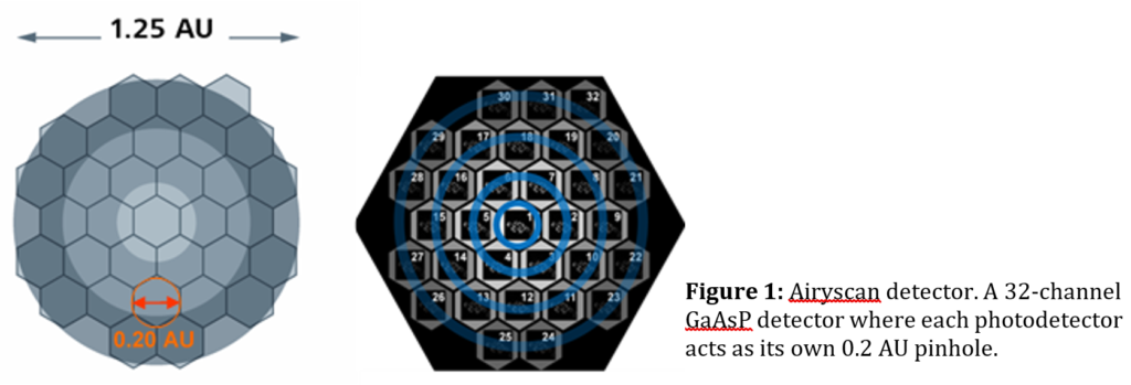
The Airyscan technique is based on traditional confocal laser scanning microscopy and is an add-on to the latest Zeiss LSM conventional confocals. The Airyscan has no physical pinhole. Instead it uses a 32 channels area detector in a honeycomb organization (see Fig. 1), with each detector acting as an individual pinhole. It is collecting a pinhole plane image for every scan with 1.25 Airy unit diameter (AU) equivalence. Each detector element acts as a small pinhole (with 0.20 Airy unit diameter) and records a complete image. The array detection of shifted pinhole picture results in less light collected per sub element of the detector but with narrower point-spread-function (PSF), i.e. increased resolution. The pixel reassignment of the shifted pinhole image increases signal-to-noise level by 4- to 8-fold compared with conventional confocal detection, which is further improved by linear deconvolution image processing.

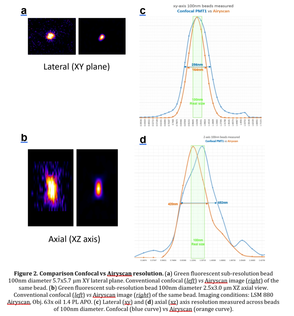
The lateral resolution (xy) goes from approx. 260 nm in conventional confocal microscopy to 140-160 nm with the Airyscan. As an additional bonus, the axial resolution (z) drops down from 600-700 nm to 400nm. Therefore you can expect a 1.7 to 1.8 times better resolution with the Airyscan detector compared with confocal detection without any real tradeoff. Fig. 2 shows improvements of resolution obtained with sub-micrometer fluorescent beads.

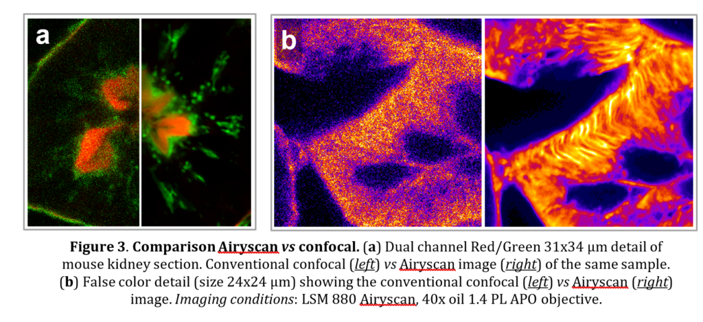
Images of biological samples are shown in Fig. 3. The increased image quality and detail in the images can be readily observed.
If you have interest, need more information or want to try this technique feel free to contact your local technical manager or Arnaud Paradis, CIF Dorigny technical Manager.
Arnaud Paradis
